
For decades, patients were told that once plaque builds up in the arteries, it’s permanent – the best you can do is stop it from getting worse. That’s no longer the full story. A growing body of high-quality imaging research now shows that with intensive treatment, artery plaque can shrink. For patients working with a concierge doctor in Las Vegas, this changes the conversation from “manage the damage” to “actively reduce it.”
To be clear from the outset: this isn’t about erasing every trace of plaque or reversing decades of buildup with a supplement. The evidence is specific about what regresses, by how much, and under what conditions. Here’s what the research actually supports, and where a concierge doctor in Las Vegas, Summerlin, or Henderson fits into making it happen.
A quick note on the evidence: the findings below come from randomized, core-lab–adjudicated imaging trials. These are some of the strongest study designs in cardiovascular medicine. This isn’t preliminary or preclinical data; it’s been replicated across multiple independent trials using serial imaging of the same arteries over time.
Is Atherosclerosis Reversible?
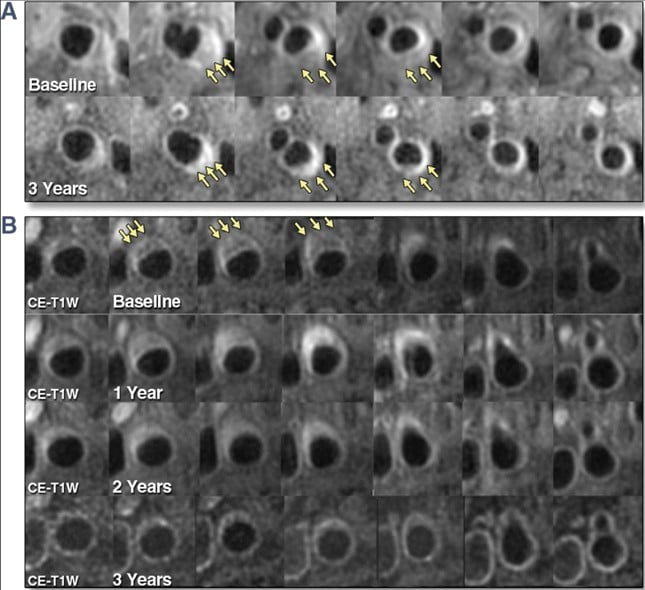
Yes – at least in part. The strongest evidence comes from trials that used serial intravascular imaging (ultrasound and optical imaging catheters threaded into coronary arteries) to measure the same plaque, in the same patient, before and after treatment. When LDL cholesterol (“bad cholesterol”) is lowered intensively enough, plaque volume measurably shrinks. Parallel studies using MRI of the carotid arteries (in the neck) confirm the same pattern, with plaque regression and favorable changes in what the plaque is made of.
The effect is dose-dependent: the lower the LDL-C achieved, the more regression occurs. And it isn’t uniform across the whole plaque – the “soft,” lipid-rich, unstable portion shrinks, while the calcified, “hard” portion tends to increase. More on why that matters below.
The Coronary Evidence: Direct Imaging Inside the Arteries
The highest-quality data comes from randomized trials using intravascular ultrasound (IVUS) and optical coherence tomography (OCT) – imaging catheters that measure plaque volume directly, inside the artery, with independent core labs reading the results.
- ASTEROID, REVERSAL, and SATURN studies established that high-intensity statins can halt, and in many patients reverse, plaque buildup. In REVERSAL, high-dose atorvastatin (80 mg) showed no significant plaque progression, while a lower-intensity statin did progress. In SATURN, both high-dose rosuvastatin and atorvastatin reduced percent atheroma volume, and the majority of patients – roughly two-thirds – showed measurable regression.
- GLAGOV, PACMAN-AMI, and HUYGENS looked at PCSK9 inhibitors, a newer class of cholesterol-lowering injectable medication, added on top of statins. These trials found further reductions in plaque burden beyond statins alone, along with thickening of the fibrous cap covering the plaque – a sign of a more stable, less rupture-prone lesion, seen roughly a year after starting treatment.
- Across every major trial, the pattern is consistent: the lower the LDL-C a patient achieves, the more the plaque regresses. Regression becomes most likely once LDL-C drops below roughly 70–80 mg/dL.
The Carotid Evidence: What Serial MRI Shows
The most direct look at plaque regression itself, not just risk of future events, comes from serial MRI of the carotid arteries.
- A prospective MRI study following patients on lipid-lowering therapy found the lipid-rich, unstable core of carotid plaque shrank progressively over time, with about 11% of patients achieving complete depletion of that unstable core over three years.
- A separate trial of the PCSK9 inhibitor alirocumab found the percentage of lipid-rich core in carotid plaque dropped substantially over 12 months, with a corresponding increase in more stable fibrous tissue.
- Longer-term MRI data (the Rotterdam study) showed that high-dose statins shift carotid plaque composition away from the unstable, lipid-heavy type and toward a more stable, calcified type – a pattern confirmed across multiple studies using carotid ultrasound, MRI, and CT angiography.
Why “Soft” Plaque Shrinks But “Hard” Plaque Doesn’t
This is the detail most people miss, and it’s the key to understanding what “reversal” actually means.
Under intensive treatment, the soft, lipid-rich, and necrotic (dead-tissue) portions of plaque shrink. But the calcified, hardened portions tend to increase in volume, not decrease. That’s not a treatment failure – it’s the imaging signature of a plaque becoming more stable. Think of it less as plaque disappearing and more as the dangerous, unstable material inside the plaque being replaced with denser, calcified, less rupture-prone material.
This matters clinically because plaque rupture, not plaque size on its own, is what triggers most heart attacks and strokes. A smaller, more stable plaque is a meaningfully lower-risk plaque, even if a coronary calcium score technically goes up.
So Your CAC Score or Carotid Ultrasound Came Back Positive, Now What?
This is often where the research above stops being abstract. Maybe your doctor ordered a carotid ultrasound because of a family history, or a Coronary Artery Calcium (CAC) test as part of a longevity or executive physical – and it came back positive. A number on a report, and suddenly “atherosclerosis” isn’t a textbook term, it’s something in your arteries right now.
A few things are worth understanding before you panic:
- A positive result is a starting point, not a verdict. A CAC score or a carotid plaque finding tells you plaque is present and roughly how much. It doesn’t tell you, on its own, whether that plaque is stable or unstable, or exactly what your risk of a heart attack or stroke looks like without more context. Your LDL-C, blood pressure, family history, and other risk factors all shape what that number means for you.
- Remember the calcium paradox from above. A rising CAC score under treatment isn’t automatically bad news. As the section above explains, effective LDL-lowering therapy can increase calcified volume while shrinking the soft, unstable plaque that actually causes events. So a higher score after starting treatment can reflect stabilization, not progression. Interpreting a follow-up scan without that context can be genuinely misleading.
- This is exactly the scenario the imaging trials were built to inform. The research above exists because people in your position need to know what to do next. A positive scan is the trigger for figuring out your target LDL-C, whether a statin alone gets you there or a PCSK9 inhibitor should be added, and how to track progress over time – not a reason to assume the damage is fixed and permanent.
- The next move is a conversation, not a Google search. A positive CAC or carotid finding raises real questions – how aggressive should treatment be, how often should this be rechecked, does this change anything else in your care plan – that deserve time with a physician who has your full history, not a five-minute checkbox visit.
If you’re holding a report like this right now, that’s precisely the kind of result a concierge doctor in Las Vegas, Summerlin, or Henderson should be walking through with you in detail, not just flagging and moving on.
How Confident Are We in This Research?
Very. The coronary IVUS/OCT trials are considered some of the strongest evidence in cardiovascular medicine: randomized, blinded, and read by independent core labs rather than the treating physician. They’re also mechanistically consistent with outcomes data – across trials, roughly every 1% reduction in plaque volume tracks with about a 20% lower odds of a major cardiac event in pooled analyses.
The carotid MRI evidence is compelling but somewhat less definitive, largely because it comes from smaller studies that often followed one group of patients over time rather than randomizing patients to different treatments.
Frequently Asked Questions
Can plaque completely disappear once it’s calcified?
No. Calcified (“hard”) plaque tends to increase in volume under treatment, not shrink. What regresses is the soft, lipid-rich, and necrotic-core material – the unstable component most likely to rupture and cause a heart attack or stroke.
What LDL level do I need to reach for plaque regression?
The trials show regression becomes most consistent once LDL-C is lowered below roughly 70–80 mg/dL. The relationship is dose-dependent – lower LDL-C generally means more regression, not a single cutoff that either works or doesn’t.
Do I need a PCSK9 inhibitor, or is a statin enough?
High-intensity statins alone produced measurable regression in multiple trials. PCSK9 inhibitors, added on top of statins, produced additional regression and favorable plaque changes in trials like GLAGOV and HUYGENS. Which approach makes sense depends on your baseline LDL-C, cardiovascular risk, and how your levels respond to a statin alone.
Is this the same as reversing heart disease?
Not entirely. This is regression of plaque volume and stabilization of plaque composition, both linked to lower cardiovascular risk. It’s not the same as eliminating existing coronary artery disease or guaranteeing you’ll never have a cardiac event.
How would I know if my own plaque is regressing?
The imaging techniques used in these trials (IVUS, OCT, serial MRI) aren’t standard outpatient tools. In practice, tracking is done through achieved LDL-C, other lipid markers, and sometimes coronary calcium scoring or CT angiography over time – which is exactly the kind of longitudinal tracking a concierge medicine model is built for.
Why This Belongs in a Conversation With Your Concierge Doctor in Las Vegas
This research has a real, practical implication: aggressive LDL-C lowering isn’t just about preventing further plaque buildup – it can actively shrink and stabilize plaque that’s already there. But translating that into an individual treatment plan means knowing your starting LDL-C, your calcium score or imaging history, your risk factors, and how aggressively to treat given your overall health picture – not applying a one-size-fits-all target.
That’s a conversation that takes time most primary care visits don’t allow. As concierge doctors serving Las Vegas, Summerlin, and Henderson, our practice has the time to review your lipid panel in detail, discuss whether a statin, a PCSK9 inhibitor, or both make sense for you, and track your progress over months and years rather than in a single annual visit.
The Bottom Line
Atherosclerosis is not a one-way street. High-quality imaging trials show that intensive LDL-C lowering – primarily with high-intensity statins, with additional benefit from PCSK9 inhibitors – produces measurable regression of coronary and carotid plaque, concentrated in the unstable, lipid-rich portions most likely to cause a heart attack or stroke. Calcified plaque tends to increase as a sign of stabilization, not treatment failure. The effect tracks closely with how low LDL-C is driven, with regression becoming most consistent below roughly 70–80 mg/dL.
Sources
Dawson, L. P., Lum, M., Nerleker, N., Nicholls, S. J., & Layland, J. (2022). Coronary atherosclerotic plaque regression: JACC state-of-the-art review. Journal of the American College of Cardiology, 79(1), 66–82. https://doi.org/10.1016/j.jacc.2021.10.035
Sarraju, A., & Nissen, S. E. (2024). Atherosclerotic plaque stabilization and regression: A review of clinical evidence. Nature Reviews Cardiology, 21(7), 487–497. https://doi.org/10.1038/s41569-023-00979-8
Ahmadi, A., & Narula, J. (2017). Primary and secondary prevention, or subclinical and clinical atherosclerosis. JACC: Cardiovascular Imaging, 10(4), 447–450. https://doi.org/10.1016/j.jcmg.2017.02.001
Zhao, X. Q., Dong, L., Hatsukami, T., Phan, B. A., Chu, B., Moore, A., Lane, T., Neradilek, M. B., Polissar, N., Monick, D., Yuan, C., & Brown, B. G. (2011). MR imaging of carotid plaque composition during lipid-lowering therapy: A prospective assessment of effect and time course. JACC: Cardiovascular Imaging, 4(9), 977–986. https://doi.org/10.1016/j.jcmg.2011.06.013
Lepor, N. E., Sun, J., Canton, G., Contreras, L., Hippe, D. S., Isquith, D. A., Kedan, I., Zhao, X. Q., & Yuan, C. (2021). Regression in carotid plaque lipid content and neovasculature with PCSK9 inhibition: A time course study. Atherosclerosis, 327, 31–38. https://doi.org/10.1016/j.atherosclerosis.2021.05.008(PMID: 34038761)
Saba, L., Saam, T., Jäger, H. R., Yuan, C., Hatsukami, T. S., Saloner, D., Wasserman, B. A., Bonati, L. H., & Wintermark, M. (2019). Imaging biomarkers of vulnerable carotid plaques for stroke risk prediction and their potential clinical implications. The Lancet Neurology, 18(6), 559–572. https://doi.org/10.1016/S1474-4422(19)30035-3
Biondi-Zoccai, G., Mastrangeli, S., Romagnoli, E., Peruzzi, M., Frati, G., Roever, L., Giordano, A., & Sciarretta, S. (2018). What we have learned from the recent meta-analyses on diagnostic methods for atherosclerotic plaque regression. Current Atherosclerosis Reports, 20(3), 15. https://doi.org/10.1007/s11883-018-0717-y
Li, Y., Deng, S., Liu, B., Yan, Y., Du, J., Li, Y., & Jing, X. (2021). The effects of lipid-lowering therapy on coronary plaque regression: A systematic review and meta-analysis. Scientific Reports, 11(1), 7999. https://doi.org/10.1038/s41598-021-87528-w